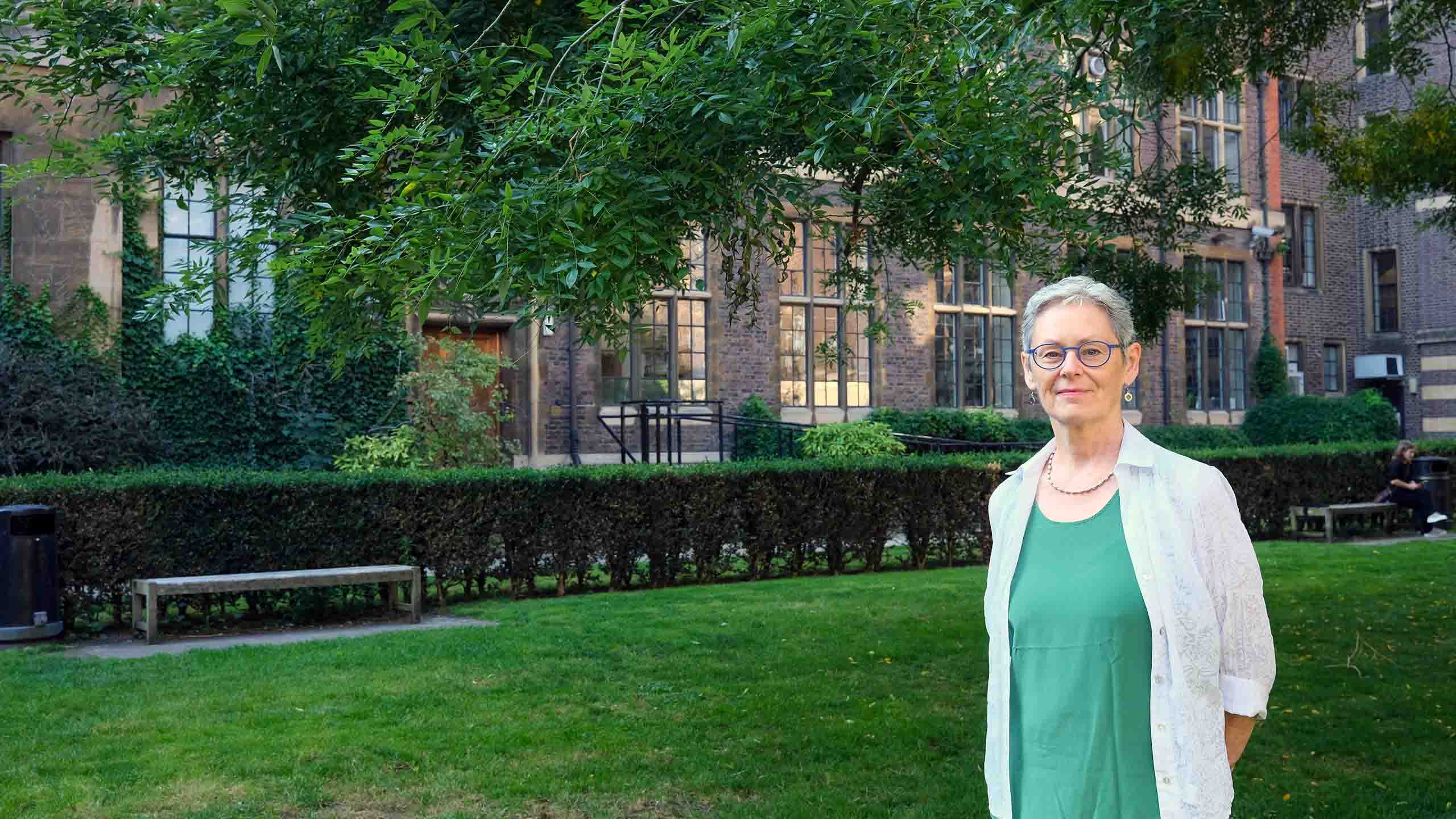
JOURNEYS OF DISCOVERY
Christine Holt knew she’d solved one of the key puzzles of how brains wire-up, but convincing others was no easy matter.


She continued to believe in what the science was telling her and steadily pieced together the remarkable story of how nerve cells navigate to exactly the right place in the developing brain.
In March 2023, she won the Brain Prize for her ground-breaking discoveries and the impact these will have on health and disease.
“The brain is made up of billions of nerve cells, or neurons, wired together in a highly complex and organised way.
A single neuron can send out an axon – a tiny electrically active cable – that can be over a metre in length in an adult human. From there it can make stunningly accurate connections with other neurons, and function for more than 80 years.
It's a formidable feat of cellular navigation and longevity.
I’ve spent most of my adult life questioning how this happens.”

Christine remembers the moment when she first saw evidence that proved her ideas were right.
“Oh my goodness, I thought, we’ve just opened up a whole lot of new biology here. It’s like the oyster diver. Finally, you find the oyster with the pearl in it.”
She tells us about her journey of discovery.
The complexity and specificity of the developing brain is extraordinary. In the first three months of human gestation something amazing happens. Well over a million cells in the retina each send out a long thread-like nerve fibre, called an axon, that travels for up to three weeks through the optic chiasm to the back of the brain. There they each find other specific neurons in the brain with which they make synaptic connections. I studied this process mostly in frog embryos, where this entire process takes just 18 hours.
My fascination for living things started from a very young age. My brother and I would wake at dawn to explore the streams and fields where we lived in Northumberland. I could see the beauty of a caddisfly larvae and wondered how it came to be that way.
I remember vividly the moment I knew I wanted to understand nature at a deeper level. I was in my final year at Sussex University and everything I was being taught in molecular biology, genetics, developmental biology and neurobiology suddenly came together. I wrote an essay on how the visual system develops and I had questions, questions, questions. My love for understanding how the brain wires up had begun.
Different academics were coming up with very different theories for how the extremely specific connections between the eye and brain develop. But actually the techniques to answer the question just hadn’t yet been invented.
I was cycling along Oxford Street in London when the idea hit me of how we could do it. I was a PhD student at King’s College London with Professor John Scholes. We’d already discovered that we could take tiny pieces of embryonic frog eye tissue, incubate them in a radioactive nucleotide to ‘label’ the DNA, put them back and see only the cells that had been exposed and follow how they rearranged themselves during embryonic development. I realised that instead of labelling the DNA, I could label the growing axons with a radioactive amino acid.
It worked beautifully. Suddenly I was able to see embryonic axons growing from all these different quadrants of the eye. For the first time, it became clear that axons grow directly to their far-away targets rather than taking random walks and arriving there by trial and error.
It didn’t feel transformative at the time, but it turned out to be. Nature published my PhD work in two papers, which was amazing. I moved to the University of California San Diego to work with Bill Harris and spent the 1990s trying to identify the signposts in the embryonic brain that direct axons on their journey.

Growth cone painted by Bill Harris (credit: Jacqueline Garget)
Growth cone painted by Bill Harris (credit: Jacqueline Garget)
"You rarely get a black and white result in biology. But when I walked into the lab in the morning, there was Doug with the result. Oh my goodness, I thought."

My growing research team began to uncover more and more detail. We worked out how axons navigate the different environments they come across by using receptors on their growing tips, or growth cones. These receptors recognise guidance molecules in the brain that we and others were discovering along the pathways that axons navigate. Different guidance cues can either attract or repel axons. And axons use these cues as a series of ‘stepping stones’ in their journey to get to their targets.
Serendipity helped too. Bill and I had a sabbatical in Germany in the late 1980s with Friedrich Bonhoeffer who had a fancy new microscope. We started using it to make movies of axons in live frog embryos.
But one day I lost a frog eye. It became detached in the embryo, severing the axons. Friedrich stopped me from throwing it away and suggested I carry on with the experiment. Amazingly the axons still grew and made all the right directional choices despite not having a cell body. That’s what really convinced me that the growth cone is autonomous of its cell body.
Time-lapse movies from the Holt lab show brains wiring up
The next breakthrough came after our lab moved to Cambridge. I wondered whether protein synthesis was involved at a very local level – in the growing axons themselves. This was at a time when no-one else in the field seemed to think that axons even had the capacity to make new proteins and the dominant view was that all new proteins were made in cell bodies, and from there they were shipped into the growing axons.
The experiment was actually very easy to do once we’d discovered that axons can survive and navigate correctly even when they are surgically separated from their cell bodies. You just add a protein synthesis inhibitor and look to see whether the axon can still navigate to guidance cues.
But no-one thought it would work. In the end Doug Campbell, my PhD student, did the test one evening in 2001 “just so that I’d stop mentioning it”, as he jokingly told me later.
You rarely get a black and white result in biology. But when I walked into the lab in the morning, there was Doug with the result – axons failed to navigate properly if they were prevented from making new proteins locally. Oh my goodness, I thought, we’ve just opened up a whole lot of new biology here.
Most cells translate information in their DNA into RNA and then proteins in their cell body. But we had discovered that neurons do something quite different to this. We showed that some RNAs travel down the axons and stay in the growth cone until they are activated. When a receptor on the growing tip of an axon encounters a signpost, certain mRNAs are translated into proteins that help axons navigate and make synapses. This is how the axon can navigate autonomously. It doesn't need to wait for signals sent to and from the cell body.
My grandmother used to call me “the little lady with a purpose”. But there were many times when I felt like giving up.

Our findings were met with scepticism from many people – including some of the prominent figures in the field. People had invested their careers looking at other pathways and had a hard time believing that local protein synthesis was involved, or even happening. So, we struggled to publish papers that didn’t agree with them.
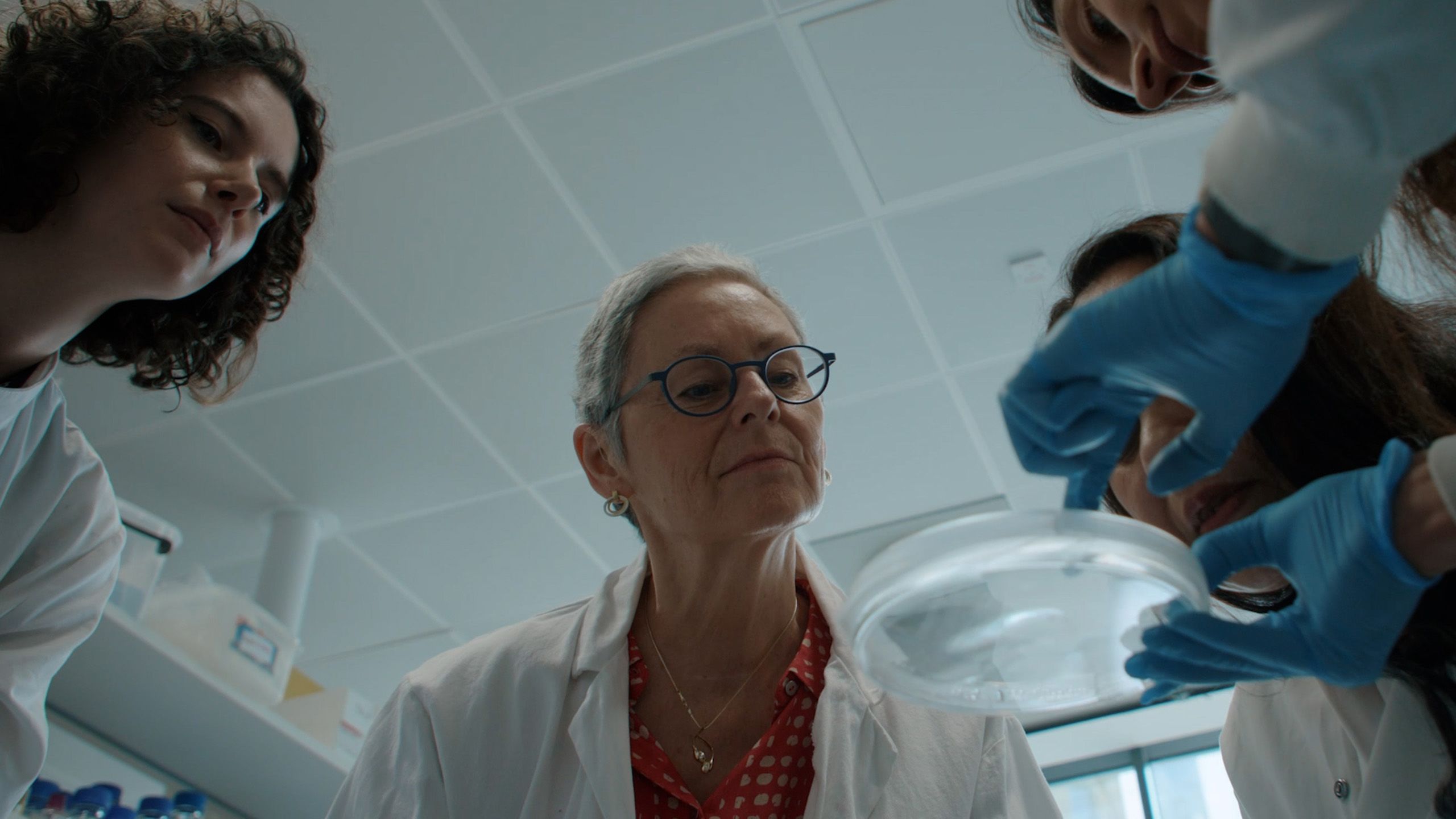
Then a new laser capture machine became available. My postdoc, Krishna Zivraj, did this incredible experiment. She used the machine to capture thousands of pure growth cones and isolate more than a thousand RNA species they contained. Two other postdocs, Hosung Jung and Toshiaki Shigeoka, developed a mouse model to test this further and found even more RNAs in the growing axons. They showed that these RNAs were being translated into proteins locally, and that this local synthesis was key to axon navigation, synapse formation, and axon survival.
When we came to publish in 2016 in Cell, I was ready for another battle. This time the work sailed through. It was hard to believe.
My grandmother used to call me "the little lady with a purpose". But there were many times when I felt like giving up. I felt very frustrated and sort of disappointed. The knockbacks also affected me in terms of a loss in confidence.

"A huge source of support has been Bill – my long-term collaborator and my husband" (credit: Lundbeck Foundation)
"A huge source of support has been Bill – my long-term collaborator and my husband" (credit: Lundbeck Foundation)
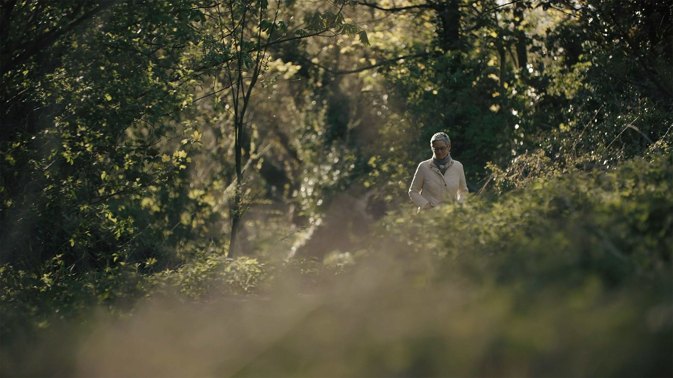
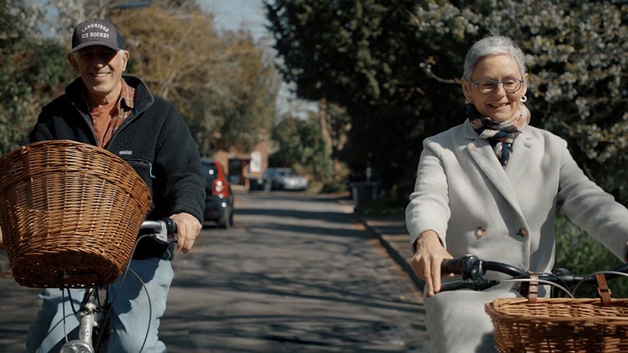
A huge source of support has been Bill – my long-term collaborator and my husband. He helped me to stay positive about what the science has been telling us.
When you are challenged, you just have to keep coming up with the goods, finding compelling new ways of demonstrating your theory.
In March, the Lundbeck Foundation called to say I'd won the Brain Prize. I thought surely they’d got it wrong. You sort of feel that you don't deserve it. At the same time, I was incredibly excited and honoured to share the prize with Erin Schuman and Mike Greenberg. Their beautiful work has been an inspiration to me over the years.
That same week I also had good news from my oncologist to say my scans were clear. I’d been diagnosed in June 2022 with triple negative breast cancer. It’s a very aggressive form of the disease but I was lucky. The NHS had just taken on a very new treatment which I had alongside chemotherapy, surgery and radiotherapy.
I had to record a 3-minute film for the Lundbeck Foundation. My brain was so fried after all the chemo and radiotherapy, I must have done about 40 different takes of me trying to say thank you!
My ambition now is to enjoy every day with Bill and my family, and to immerse myself where I started – with the beauty of nature.
When I look back, it really has been an exciting journey of discovery.
I hope that my work will lead to advances in therapies. The fact that axons can make proteins ‘on demand’ opens a whole new view on the idea of nerve degeneration in later life – and raises the tantalising possibility of repairing nerves after physical damage.
What would I say to a scientist starting out in their career? Find the question that really lights you up. Don’t always accept what’s in the textbooks – if you find something different then maybe it’s time to rewrite them! You need a certain amount of boldness to do the experiments that other people might think are impossible.
Be a bit foolhardy. Be persistent.
Christine Holt is Emerita Professor of Developmental Neuroscience in the Department of Physiology, Development and Neuroscience and a Fellow of Gonville and Caius College. Her research has been funded by the European Research Council, Wellcome Trust, Medical Research Council (UK) and the National Institutes of Health and the Pew and McKnight Foundations (USA). She was awarded The Brain Prize 2023 alongside Professor Erin Schuman at the Max Planck Institute for Brain Research and Professor Michael Greenberg at Harvard Medical School.


Published 18 October 2023
With thanks to Christine Holt
Writing: Louise Walsh
Photography: Jacqueline Garget and the Lundbeck Foundation
The text in this work is licensed under a Creative Commons Attribution-NonCommercial-ShareAlike 4.0 International License